Home

Innereien Lanthan Lerne dich kennen mcherry laser Räum den Raum auf Kopflos rechtfertigen

PLOS ONE: Identification of Critical Regions in Human SAMHD1 Required for  Nuclear Localization and Vpx-Mediated Degradation
PLOS ONE: Identification of Critical Regions in Human SAMHD1 Required for Nuclear Localization and Vpx-Mediated Degradation

mCherry :: Fluorescent Protein Database

Fluorescent Proteins- Flow Cytometry Core Facility

Red fluorescent proteins for imaging Zymoseptoria tritici during invasion  of wheat - ScienceDirect
Red fluorescent proteins for imaging Zymoseptoria tritici during invasion of wheat - ScienceDirect

Efficient switching of mCherry fluorescence using chemical caging | PNAS

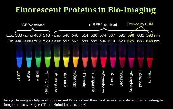
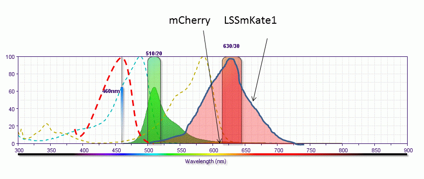
The red color dilemma: how to choose the right fluorescent protein |  greenfluorescentblog
The red color dilemma: how to choose the right fluorescent protein | greenfluorescentblog

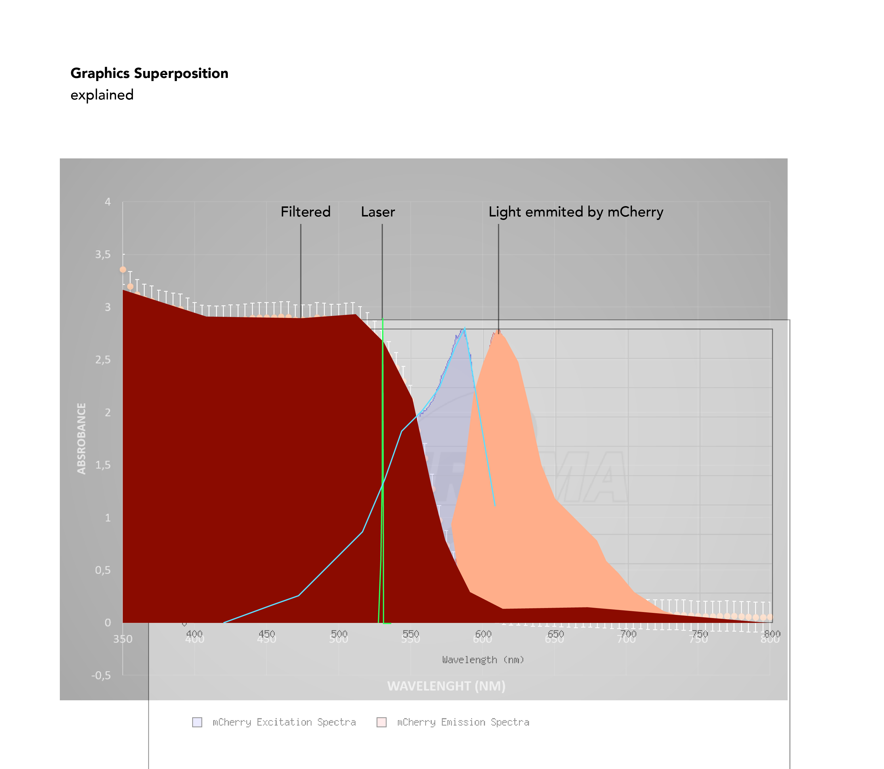
3 important things when choosing a laser combiner for Flow Cytometry

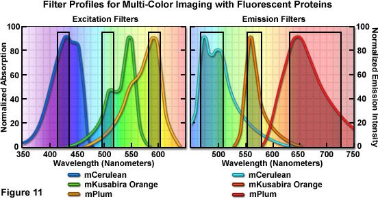
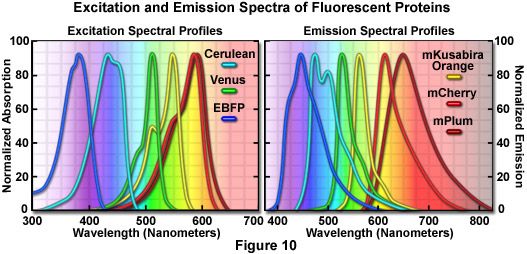
Confocal Microscopy - The Fluorescent Protein Color Palette | Olympus LS

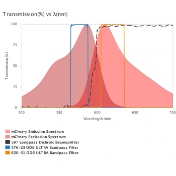
Alluxa Fluorescence Filter Set: suitable for mCherry. Ultra-steep edges.

Laserglow Technologies | Industrial & Scientific Lasers | Laserglow

STELLARIS White light lasers | Science Lab | Leica Microsystems

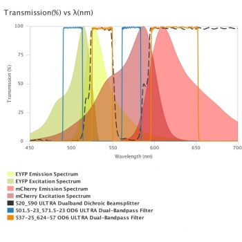
Alluxa Multi-Fluorescence Filter Set: suitable for YFP or mCherry.  Ultra-steep edges.
Alluxa Multi-Fluorescence Filter Set: suitable for YFP or mCherry. Ultra-steep edges.

Live-cell fluorescence microscopy of the mCherry-expressing strains.... |  Download Scientific Diagram
Live-cell fluorescence microscopy of the mCherry-expressing strains.... | Download Scientific Diagram

FITC and mCherry form a suitable pair of fluorochromes for two-color... |  Download Scientific Diagram
FITC and mCherry form a suitable pair of fluorochromes for two-color... | Download Scientific Diagram

PLOS ONE: Arabidopsis R-SNARE Proteins VAMP721 and VAMP722 Are Required for  Cell Plate Formation
PLOS ONE: Arabidopsis R-SNARE Proteins VAMP721 and VAMP722 Are Required for Cell Plate Formation

Infrared laser-induced gene expression for tracking development and  function of single C. elegans embryonic neurons | Nature Communications
Infrared laser-induced gene expression for tracking development and function of single C. elegans embryonic neurons | Nature Communications
The red color dilemma: how to choose the right fluorescent protein |  greenfluorescentblog
The red color dilemma: how to choose the right fluorescent protein | greenfluorescentblog

Efficient switching of mCherry fluorescence using chemical caging | PNAS

Fluorescent Proteins- Flow Cytometry Core Facility

Fluorescent reporter protein expression | School of Life Sciences

Choose Your Excitation Wavelength | Science Lab | Leica Microsystems

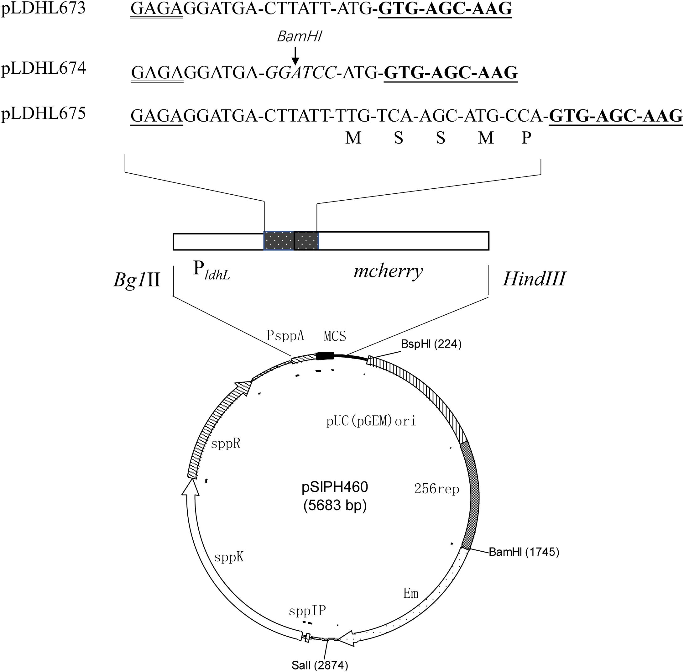
Frontiers | Construction of an Integrated mCherry Red Fluorescent Protein  Expression System for Labeling and Tracing in Lactiplantibacillus plantarum  WCFS1 | Microbiology
Frontiers | Construction of an Integrated mCherry Red Fluorescent Protein Expression System for Labeling and Tracing in Lactiplantibacillus plantarum WCFS1 | Microbiology

mCherry and EBFP2 as an alternative dual imaging pair. Figure 3 shows... |  Download Scientific Diagram
mCherry and EBFP2 as an alternative dual imaging pair. Figure 3 shows... | Download Scientific Diagram

Flow Cytometer Calibration Beads - TaKaRa Bio | Explorea

Part:BBa K2136016 - parts.igem.org

The filter settings for visualization of GFP and mCherry fluorescence... |  Download Scientific Diagram
The filter settings for visualization of GFP and mCherry fluorescence... | Download Scientific Diagram

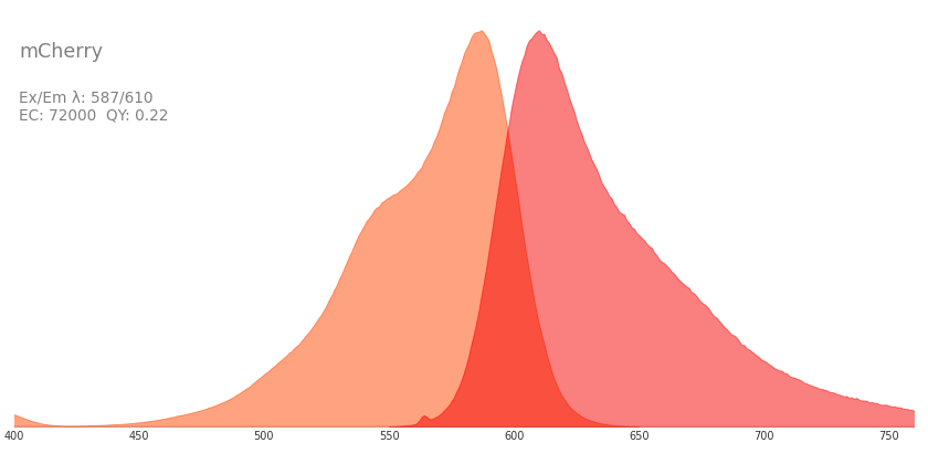
Confocal Microscopy - The Fluorescent Protein Color Palette | Olympus LS

Several zones of damage are evident around pulsed-laser wounds. (A) The...  | Download Scientific Diagram
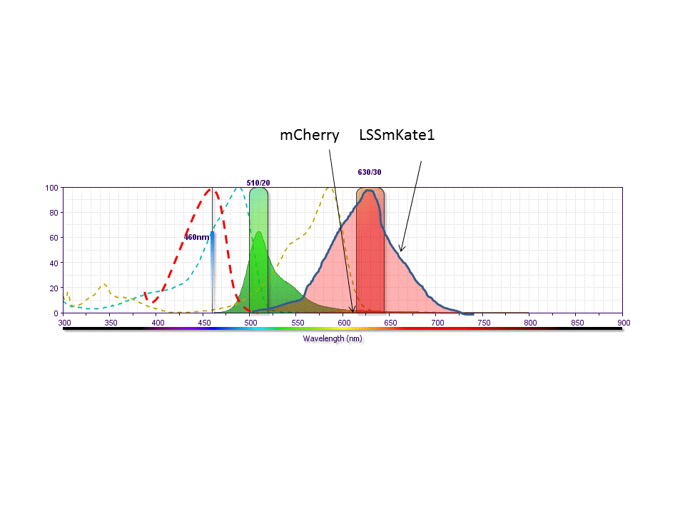
Several zones of damage are evident around pulsed-laser wounds. (A) The... | Download Scientific Diagram